A Hemispherectomy And The Postoperative Period
A hemispherectomy is a surgical procedure used to treat a variety of seizure disorders. Most often, doctors perform it when these conditions do not respond adequately to other, less radical treatments.
The first hemispherectomy was performed on a dog in 1888. However, the first reference to this procedure in humans dates back to 1923. In the 1960s and 1970s, several interventions yielded poor results.
Today this has all changed. Doctors often opt for an anatomical hemispherectomy rather than a functional hemispherectomy. This is a more accurate and less invasive intervention. The success rate is also much higher than in the past.
In this article we will take a closer look at the modern forms of this procedure.
What is a hemispherectomy?
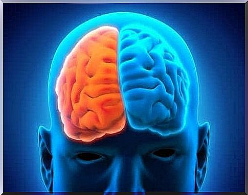
A hemispherectomy is a neurosurgical procedure in which one of the hemispheres of the brain is removed. Sometimes the surgeon removes the left hemisphere of the brain, in other cases just the right hemisphere. This depends on where the problems are.
Doctors perform this procedure mainly in extreme cases and in children aged 5 to 10 years. This type of intervention is primarily an anticonvulsant treatment. It is also useful for patients with neurological deficits and in severe cases of severe head injury.
In most interventions, the entire hemisphere of the brain is removed. Sometimes, however, only part of it is removed. This is called a functional hemispherectomy. In such cases, if the surgeon leaves a small amount of damaged tissue, the seizures may re-appear.
Indications to do a hemispherectomy
In general, medical specialists may indicate to perform this procedure on patients who suffer from continuous and daily seizures and who have not responded to other medical treatments or other less invasive surgical procedures.
Medical specialists recommend it in the following cases:
- Children with hemiplegia. This is a half-sided paralysis and the procedure is only for children older than four years who suffer from epileptic and/or psychological disorders. The patient has had checkups for two years and is not responding to drug treatment.
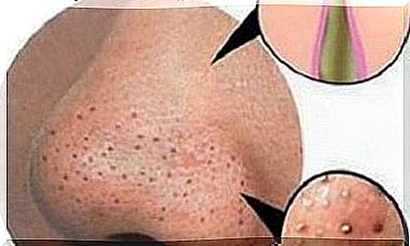
- Sturge-Weber syndrome. This is a neuro-cutaneous condition characterized by a port-wine stain on the face, in the area of the trigeminal nerve. Doctors may recommend this type of surgery when problems start at a young age and the condition affects the entire hemisphere of the brain.
- Rasmussen encephalitis. This condition causes chronic, progressive encephalitis, an inflammation of the brain. In this case, it is best to intervene early.
- Hemimegalencephaly (HME). This is a rare neurological inflammatory disease that also causes severe seizures. However, medical specialists still disagree about whether surgery is the best option for patients with this condition.
- Cortical developmental malformations.
Characteristics of the procedure
In general, there are four types of hemispherectomy. The four types are:
- Anatomical Hemispherectomy
- Hemidecortication
- Functional Hemispherectomy
- Modified Functional Hemispherectomy
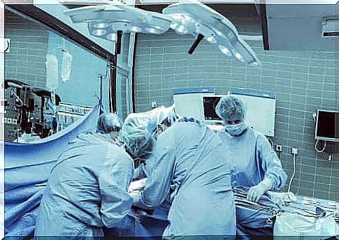
Usually, doctors use anesthesia for the procedure.
- The surgeon first begins by shaving the patient’s head and marking the incision lines.
- Then the surgeon cuts to expose the hard skull bone. He then removes these to reach the brain.
- Then he carefully marks the area he needs to remove. After removal, the blood vessels should be cauterized.
- A drainage device is then placed.
- Finally , he reinserts the skull bone and scalp and closes the incision with staples or sutures.
The postoperative period
Unfortunately, the postoperative period of this procedure is painful. Usually doctors leave the drain in place for 3 to 4 days. After that, the attending physician evaluates the situation and decides whether the tube can be removed or not. Before they remove it, he’ll need to run diagnostic tests to determine if there’s any bleeding or bruising.
However, doctors normally control and prevent the complications very well. The main complications are:
- hemodynamic instability
- hypothermia
- hypo- or hyperkalaemia
Seizures during the postoperative period are another serious complication. About half of patients develop hydrocephalus. Almost all patients develop aseptic meningitis. There is also evidence that some complications may manifest later.
Nevertheless, the death rate is quite low and varies between 4% and 6%. Between 70% and 85% of patients who undergo a hemispherectomy no longer suffer from seizures. Meanwhile, in about 10-20%, the quality of life improves significantly.